Introduction to Animal Cells for 7th Grade: 7th Grade Animal Cell Coloring

7th grade animal cell coloring – Animal cells are the fundamental building blocks of animals, including humans. They are eukaryotic cells, meaning they possess a membrane-bound nucleus and other specialized internal structures called organelles. Understanding the structure and function of these organelles is crucial to grasping how animals grow, develop, and maintain themselves. This section will explore the basic components of an animal cell and their individual roles.Animal cells, unlike plant cells, lack a rigid cell wall and chloroplasts.
Instead, their structure is defined by a flexible cell membrane and the intricate arrangement of their internal organelles. These organelles work together in a coordinated manner to carry out essential life processes.
Cell Membrane Structure and Function
The cell membrane, also known as the plasma membrane, is a selectively permeable barrier that surrounds the entire cell. It’s composed primarily of a phospholipid bilayer, a double layer of phospholipid molecules. Each phospholipid molecule has a hydrophilic (water-loving) head and two hydrophobic (water-fearing) tails. This arrangement creates a barrier that separates the cell’s internal environment from its external surroundings.
Embedded within this bilayer are various proteins that perform a variety of functions, including transporting molecules across the membrane, receiving signals from other cells, and anchoring the cytoskeleton. The cell membrane’s selective permeability allows it to regulate the passage of substances into and out of the cell, ensuring the cell maintains a stable internal environment. This regulation is crucial for maintaining proper cell function and preventing damage.
For example, the cell membrane controls the entry of nutrients and the exit of waste products. Ions, such as sodium and potassium, are also carefully regulated by the membrane’s transport proteins to maintain proper electrical potential across the membrane, essential for nerve impulse transmission.
Organelles and Their Functions, 7th grade animal cell coloring
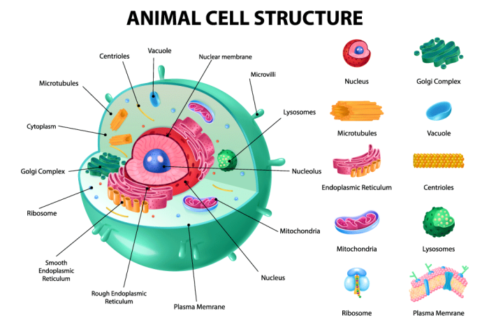
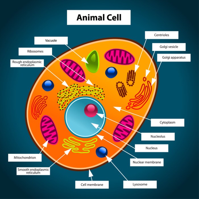
The cytoplasm, the jelly-like substance filling the cell, houses various organelles each with specific roles. These organelles work together to maintain cell life.The nucleus, the largest organelle, contains the cell’s genetic material (DNA) organized into chromosomes. It controls the cell’s activities by directing protein synthesis. The DNA’s instructions are transcribed into RNA, which then carries the instructions to ribosomes.Ribosomes are responsible for protein synthesis, translating the RNA code into amino acid sequences that fold into functional proteins.
These proteins perform a vast array of functions within the cell and the body as a whole.The endoplasmic reticulum (ER) is a network of membranes involved in protein and lipid synthesis and transport. The rough ER, studded with ribosomes, synthesizes proteins, while the smooth ER synthesizes lipids and detoxifies harmful substances.The Golgi apparatus processes and packages proteins and lipids received from the ER, preparing them for transport to other parts of the cell or secretion outside the cell.
Think of it as the cell’s post office.Mitochondria are often called the “powerhouses” of the cell because they generate energy (ATP) through cellular respiration. This energy fuels all cellular activities.Lysosomes contain enzymes that break down waste materials and cellular debris. They are essential for maintaining cellular cleanliness and preventing the buildup of harmful substances.The cytoskeleton, a network of protein filaments, provides structural support and helps maintain cell shape.
It also plays a role in cell movement and intracellular transport.
Cell Coloring Activity Design

This activity aims to reinforce 7th-grade students’ understanding of animal cell structure and function through a visually engaging coloring exercise. The design emphasizes accurate representation of organelles and their relative sizes within the cell, promoting a deeper comprehension of their roles. Color selection is guided by both visual appeal and functional relevance, enhancing memorability and understanding.The coloring page will depict a typical animal cell, featuring a variety of organelles clearly labeled.
Students will color each organelle according to a provided key, allowing for both creativity and accurate representation of cellular components. The instructions will include tips to ensure neatness and accuracy, fostering attention to detail and scientific precision.
Organelle Color Choices and Rationale
The selection of colors for each organelle is designed to aid in memorization and to reflect their visual properties or functional roles where applicable. For example, the cell membrane, a selectively permeable barrier, might be represented in a light blue to visually represent its thin and somewhat transparent nature. In contrast, the nucleus, the control center containing the genetic material, could be colored a bold, dark red to emphasize its importance.
This approach uses color as a mnemonic device to improve retention of organelle names and functions.
| Organelle | Suggested Color | Rationale |
|---|---|---|
| Cell Membrane | Light Blue | Represents its thin and somewhat transparent nature. |
| Cytoplasm | Pale Yellow | Suggests a fluid-like consistency. |
| Nucleus | Dark Red | Highlights its central role and importance. |
| Nucleolus | Darker Shade of Red | Indicates its location within the nucleus and its role in ribosome production. |
| Ribosomes | Purple | Distinguishes them as the protein synthesis sites. |
| Rough Endoplasmic Reticulum (RER) | Light Green | Visually represents the ribosomes attached to its surface. |
| Smooth Endoplasmic Reticulum (SER) | Lighter Green | Differentiates it from the RER while maintaining a similar visual theme. |
| Golgi Apparatus | Orange | Represents its layered structure and role in packaging and secretion. |
| Mitochondria | Deep Pink/Magenta | Reflects their role as the “powerhouses” of the cell. |
| Lysosomes | Dark Purple | Represents their function in waste breakdown. |
| Centrioles | Brown | Distinguishes them as involved in cell division. |
Instructions and Tips for the Coloring Activity
Prior to beginning the coloring activity, a brief review of the animal cell’s organelles and their functions will be beneficial. Students should be instructed to use crayons, colored pencils, or markers, selecting colors from the provided key. Emphasis should be placed on using the chosen colors accurately and consistently, ensuring that each organelle is clearly defined and distinctly colored.Students should be encouraged to color neatly, staying within the lines of each organelle.
This promotes careful observation and precise representation of the cell’s structure. Accurate labeling of each organelle is also crucial. Neat handwriting and clear labeling will improve the overall quality of the completed work. The activity will assess both the accuracy of the coloring and the understanding of the organelles’ functions, thus combining artistic expression with scientific accuracy.
Understanding the intricacies of 7th grade animal cell coloring requires a detailed approach, moving beyond simple diagrams. To enhance comprehension of diverse cell structures, consider supplementing your studies with visually engaging resources, such as the free printable ocean animal coloring pages , which can aid in developing fine motor skills and observation techniques applicable to microscopic cell structures.
This visual learning approach can ultimately improve your understanding of animal cell components and their functions.


